A Tiny Cluster of Brainstem Cells May Explain Why Anxiety Outlasts Its Trigger
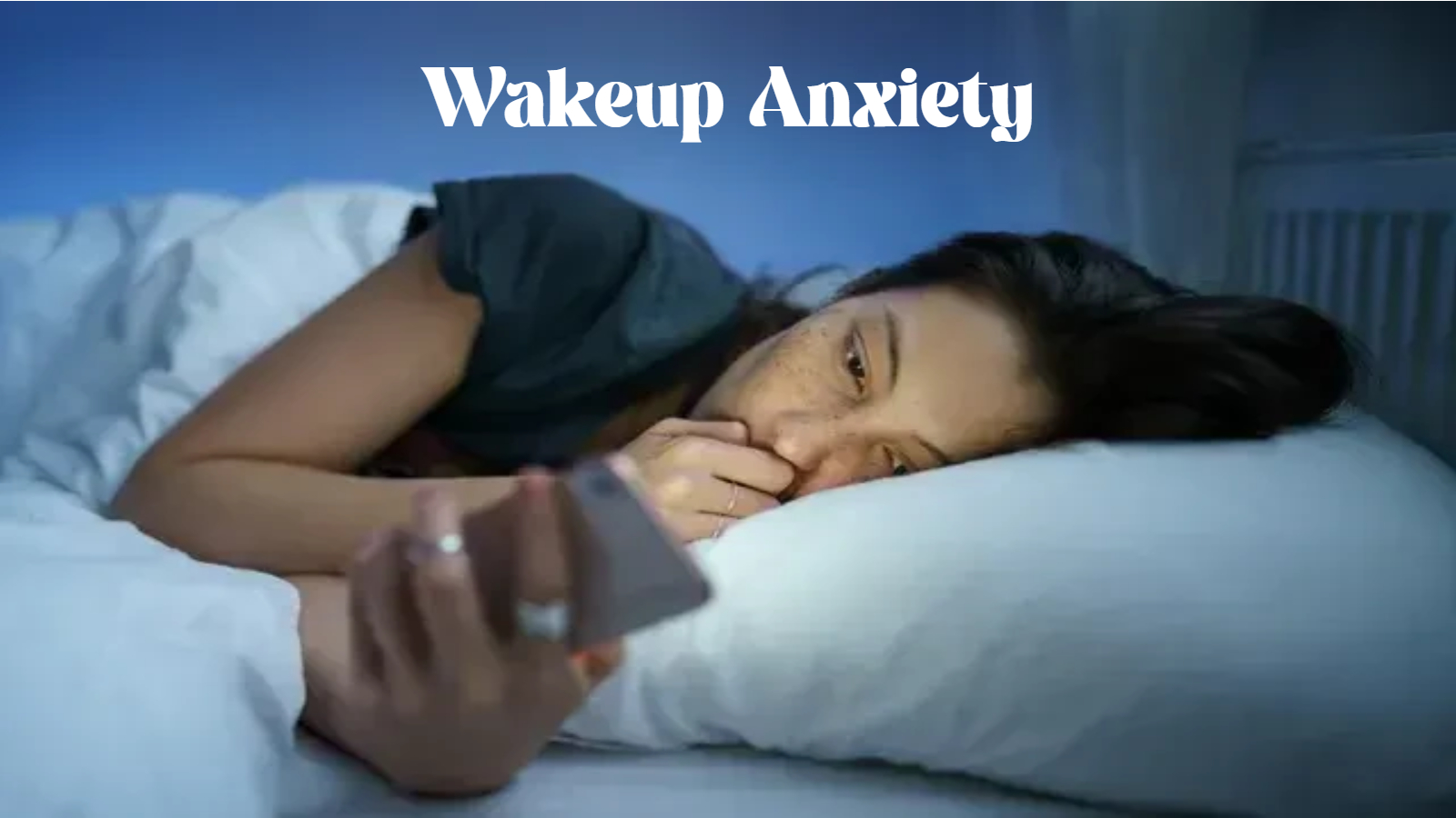
C1 neurons keep a stress hub locked “on” for days after a threat passes, a pattern that mirrors the hyper-aroused brainwaves alpha-theta training is built to calm.
A client of mine once described her anxiety as a car alarm that wouldn’t shut off. The threat was long gone. Somebody had already checked the parking lot twice. But the siren kept wailing anyway, and no amount of reasoning with it made any difference. She wasn’t being dramatic. She was describing, with more precision than she knew, exactly what a team at St. Jude Children’s Research Hospital just confirmed in a mouse model published in the journal Neuron.
For decades, the story of anxiety belonged to the amygdala and the prefrontal cortex. Those are the brain’s thinking centers, the parts we picture lighting up on a scan while someone stares down a threat. The lower brainstem got assigned the boring job: keep the heart beating, keep the lungs going, stay out of the emotional conversation. Researchers led by Lindsay Schwarz, PhD, just overturned that assumption in a fairly dramatic way, and the full findings, published in Neuron, point to a tiny cluster of cells in the medulla as a previously unrecognized master switch for fear.
A Brief Alarm That Forgets to Turn Off
The cells in question are called C1 neurons, and they live in the rostral ventrolateral medulla, a region so deep in the brainstem that it barely made it into anxiety research at all. Under normal circumstances, C1 neurons produce a short burst of epinephrine when something stressful happens. Heart rate climbs, attention sharpens, the body braces. Then, once the threat passes, the alarm resets.
The St. Jude team found that <cite index=”3-1″>strong activation of these neurons may keep that alarm switched on far longer than it should stay on, producing anxiety that persists for days</cite>. Mice with heavily activated C1 circuits showed elevated anxiety-like behavior that lingered for roughly a week after a single stressful trigger, long after the actual stressor had ended. The mechanism runs through a downstream hub called the periaqueductal gray, or PAG, which the C1 neurons excite directly. Once that hub gets locked into an overactive state, it keeps broadcasting threat signals even without new input.
<cite index=”6-1″>C1 neurons appear to drive anxiety without touching the basic autonomic processes the brainstem normally handles</cite>, which is the detail that has researchers excited. Most existing anxiety medications work by dampening signaling broadly across the brain and body, which is part of why side effects and discontinuation rates run so high. A target this narrow, tucked into a region nobody thought to look at, opens the door to treatments that quiet the anxiety loop without touching everything else a person needs to function.
Why the Brainstem Was the Blind Spot
Ask most clinicians where anxiety lives and you’ll get variations on the same answer: amygdala hyperactivity, weakened prefrontal regulation, maybe some hippocampal involvement if they’re being thorough. That framework isn’t wrong. It’s just incomplete, and it has been incomplete for a long time because the brainstem got written off as pure autopilot.
The RVLM, where C1 neurons sit, controls blood pressure, breathing rate, and glucose regulation. It’s ancient tissue in evolutionary terms, present in some form across a huge range of vertebrate species. Finding a circuit inside that machinery capable of dictating a week-long emotional state is the kind of result that makes people go back and question their org chart of the brain. <cite index=”7-1″>The RVLM contains two subpopulations of catecholaminergic neurons known as A1 and C1 cells, and it turns out the C1 population enhances excitatory activity in the ventrolateral PAG to promote anxiety-like behavior</cite>, a connection nobody had mapped with this level of precision before.
<cite index=”7-1″>Inhibiting the C1 neurons had the opposite effect, reducing anxiety and dampening the excitatory response in the PAG</cite>, which gives the finding real therapeutic weight rather than just academic interest. If you can turn the switch down, you can potentially turn a chronic anxiety state back off.
Where EEG Hyperarousal Fits Into the Picture
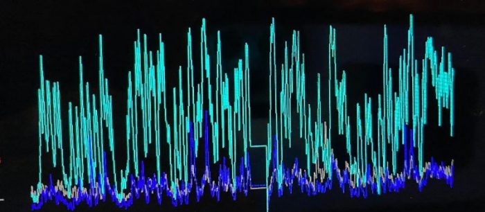
Here’s where this research starts to rhyme with something we watch on the scalp every single day in the clinic. Anxious brains have a signature, and it shows up reliably on quantitative EEG. <cite index=”14-1″>Patients with generalized anxiety disorder consistently show increased beta-band activity, and this elevated beta pattern reflects an abnormal hyperarousal of brain alertness</cite>. The frontal cortex gets stuck producing fast, high-frequency waves in the 20 to 30 Hz range, and clinically that pattern tracks with rumination, hypervigilance, and a nervous system that can’t find its off switch.
The parallel to C1 neurons is hard to miss. Both describe a system that overshoots its own reset. The brainstem circuit floods the PAG with excitatory signal long after the original threat is gone. The cortex, meanwhile, keeps generating high-beta activity that mirrors that same locked-on state, one level up in the nervous system. Whether the cortical hyperarousal is downstream of PAG output, a parallel expression of the same dysregulated stress axis, or some combination of both is still an open question that will need more direct human research to answer. But clinically, we don’t need to wait on that mechanism to be fully mapped to see the pattern show up on a screen.
I’ve watched this play out across thousands of client sessions over 17 years. Someone comes in describing exactly the car-alarm feeling my client described, and the quantitative EEG shows frontal beta running hot, often paired with suppressed alpha, the brain’s idle frequency. The nervous system isn’t choosing to stay alert. It’s stuck there, the same way those mice stayed anxious for a week after their C1 neurons fired too hard. Bottom-up dysregulation, whether it starts in the brainstem or gets amplified by it, produces a cortical signature we can actually see and, more importantly, actually train.
Alpha-Theta Neurofeedback at Sleep Recovery
Alpha-theta training earns its place in anxiety and trauma work in exactly this territory. The protocol guides the brain toward the alpha-theta crossover, a deeply relaxed state where alpha waves (8 to 12 Hz) and theta waves (4 to 8 Hz) begin to trade dominance. It’s the same threshold the brain passes through on the way into sleep, and it turns out to be a remarkably fertile place to retrain a nervous system that’s been stuck in high gear.
At Sleep Recovery, alpha-theta sessions are built around real-time EEG feedback rather than guesswork. A client settles into a reclined, low-stimulation setup while sensors track brainwave activity continuously. When the brain produces the target alpha-theta ratio, it gets an immediate auditory cue, usually a soft tone or shift in ambient sound. No conscious effort is required and none is particularly helpful. The learning happens implicitly, the same way a body learns balance on a bicycle rather than through a lecture on physics.
What makes this relevant to the C1 neuron findings is the direction of the training itself. Where a hyper=aroused nervous system needs a circuit breaker, alpha-theta training gives the cortex thousands of small opportunities, session after session, to practice dropping out of high-beta and into a slower, calmer rhythm. Clients with trauma histories often describe the sessions as the first time their nervous system seemed to remember how to actually power down rather than just distract itself into temporary quiet.
We also pair alpha-theta work with baseline quantitative EEG mapping before starting a protocol. Two clients can walk in with the same anxiety complaint and show completely different brainwave signatures, one with frontal beta hyperarousal, another with more of a suppressed alpha and asymmetry pattern, and the training gets tailored accordingly. That precision matters. Generic relaxation techniques help some people and do almost nothing for others, largely because they aren’t matched to what the brain is actually doing.
None of this depends on the C1 neuron findings being replicated in humans to be useful today. The EEG hyperarousal pattern is already well documented, the alpha-theta protocol already has a research history stretching back decades, and clients don’t need to wait on a mouse study to get relief from a nervous system that won’t quiet down. But it’s worth noting how well the two lines of evidence line up. A deep, ancient brainstem circuit gets stuck in the “on” position and the cortex it feeds shows exactly the kind of locked-in hyperarousal we spend our clinical days trying to untrain.
Sessions typically run in a series, not a single visit, because the nervous system needs repetition to build a new default rather than a temporary state. Most clients start noticing changes in sleep onset and general reactivity within the first several sessions, well before the full course finishes. Better sleep and lower anxiety tend to move together anyway. A brainstem stuck signaling threat doesn’t just produce daytime hypervigilance. It also keeps the nervous system from settling into the deeper stages of sleep where actual physical and emotional repair happens, which is part of why so many anxiety clients arrive at Sleep Recovery already exhausted on top of anxious.
There’s also a trauma-specific version of this training that goes a step further than standard alpha-theta protocols. Clients with a documented trauma history sometimes move through what clinicians call an abreactive phase during sessions, where old material surfaces briefly as the brain drops into that deep alpha-theta state. It can feel unsettling in the moment, but it tends to resolve within the session itself, and clients often describe a sense of lightness afterward that outlasts the appointment by days. We screen carefully for who is a good candidate for this deeper protocol versus a more gradual approach, since not every nervous system is ready to process that material the same way or at the same pace.
What This Means Going Forward
<cite index=”1-1″>The trial revealed that over activating this circuit permanently alters downstream PAG signaling, causing severe anxiety behaviors that persist for a full week after the initial trigger</cite>, and that single sentence probably describes more human anxiety experiences than most of us realize. A single bad night, a single frightening event, a single flood of stress hormones, and the nervous system stays braced for a threat that already passed.
Drug development aimed at C1 neurons is still early, confined to animal models, and years from clinical availability if it pans out at all. Neurofeedback isn’t waiting on that timeline. It works with the signal the brain is already producing, right now, on the same afternoon someone walks through the door. Anxiety disorders affecting more than 300 million people worldwide deserve every available angle, from brainstem pharmacology decades out to EEG-guided training available this week.
If a nervous system stuck in overdrive sounds familiar, alpha-theta neurofeedback is worth a real conversation, not just another relaxation app.
Sleep Recovery, Inc. Neurofeedback and brainwave entrainment for anxiety, trauma, and insomnia.